The Divisions of the Peripheral Nervous System
The nervous system consists of two major branches: the central nervous system (CNS) and the peripheral nervous system (PNS). The CNS consists of the brain and spinal cord. The peripheral nervous system consists of all the nerves on the periphery(outside) of the CNS. The PNS consists of two primary divisions: the somatic nervous system and the autonomic nervous system.
The somatic nervous system (SNS) controls your skeletal muscles. The SNS is the only division of the PNS that is under voluntary control. For example, I am in conscious control of my fingers while writing this chapter. However, skeletal muscle reflexes are involuntary. The skeletal muscle that is the diaphragm is under conscious (speech) and unconscious (respiration)control. We will explore the SNS is in more depth in the movement unit.
The autonomic nervous system (ANS) consists of two subdivisions: the sympathetic nervous system (SyNS) and the parasympathetic nervous system (PyNS).
The SyNS handles the fight-or-flight response and energy expenditure. Activation of our SyNSincreases heart rate, respiratory rate, blood flow to the limbs, and reflexes. Also, the pupils dilate, and the senses are more responsive to a person’s surroundings.
The PyNS tells the body to conserve energy. Parasympathetic responses are in opposition to sympathetic responses— i.e., slow heart rate and respiration. Also, the extra blood in the limbs diverts to the digestive system.
Both branches of the ANS are involuntary. You have no direct control over your heart rate, pupil diameter, digestion, blood flow, and the fight-or-flight response.

The Spinal Nerves
A spinal nerve is a cluster of sensory neuron axons or motor neuron axons wrapped in a fibrous sheet. The motor neuron’s cell body is in the spinal cord, and its axon extends the distance to its target organ. For example, the longest motor axon in the PNS spans the distance between the vertebral column base to the big toe.

A sensory neuron’s cell body is in a swelling near the spinal cord called the dorsal root ganglion. A ganglion is a cluster of cell bodies. The longest axon in the body is a sensory neuron that originates in the foot and travels to the brainstem.
The Cranial Nerves
The cranial nerves (CN) originate in the cranium, hence their name. The twelve cranial nerves form sensory pathways and motor pathways. The location on the brainstem indicates a cranial nerve’s number. For example, the olfactory nerve (CN #1) is the superior cranial nerve, and the hypoglossal nerve (CN #12) is the inferior cranial nerve.


Cranial Nerves Mnemonic
To learn the order of the cranial nerves, remember this phrase:
- Oh, oh, oh, to touch and feel very good velvet, ah heaven
Let’s explain this mnemonic:
- Oh = olfactory nerve
- oh = optic nerve
- oh = oculomotor nerve
- to = trochlear nerve
- touch = trigeminal nerve
- and = abducens nerve
- feel = facial nerve
- very = vestibulocochlear nerve
- good = glossopharyngeal nerve
- velvet = vagus nerve
- ah = accessory nerve
- heaven = hypoglossal nerve
There are other mnemonics out there, so you can search for those or create you own.
Here’s one I just thought of:
- Orange origami octopus’ tip toe always furry velociraptor great victory, alligator Harold
It’s a work in progress.
Cranial Nerve #1
The olfactory nerve is the first cranial nerve (CN #1) and consists of sensory neurons that send olfactory information (scents) to the brain.
Within our nasal cavities, there is a group of neurons immersed in mucus. On the neuron’s membranes are chemoreceptor proteins that are responsive to odors (chemicals). Chemoreceptors only function when odors dissolve in liquid. Mucus, a viscous liquid, bathes the olfactory receptors. Chemoreceptors’ moisture need is why smells are more pronounced in humid environments.
The olfactory nerve is the only sensory nerve with a direct, unfiltered link to our emotions, which is why the smell of a dead rat will invoke a more nauseating emotional response than the sight of the rat.

Here is a simple test to see if your olfactory nerve is doing its job. It ends with a little bit of English dry wit.

Cranial Nerve #2
The optic nerve is the second cranial nerve (CN #2) and extends from the eye’s retina to the posterior region of the brain.
Vision is our most complex sense, and the eye is the most complex sensory organ. The eye’s primary job is to focus light on the sensory neurons within the retina. Light enters the eye through the cornea, which refracts the light towards the lens of the eye. The cornea changes shape to focus the image on the retina, a process called accommodation.
Accommodation of the lens is why we can focus on something up close and far away. When an object moves within 20 feet of our sight, there is a medial movement of the eyes, and the pupils constrict. Suspensory ligaments attach the lens to the internal muscular wall of the anterior eye (ciliary muscles). The contraction and relaxation of the ciliary muscles change the shape of the lens. When an object is farther than 20 feet from our line of sight, our pupils dilate, and the eyes move laterally. The ciliary muscles relax and pull on the suspensory ligaments stretching the lens. The elongated lens lets us see objects in the distance.

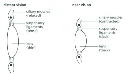
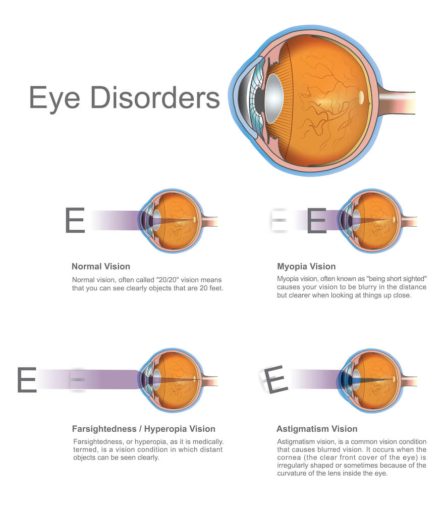

The near point is the closest distance an object can get to your eye without losing focus. The average near point for a senior in high school is 25 centimeters (one inch). However, our lens thickens throughout our life. The thicker the lens gets, the less able it is to focus on near objects. Around the age of forty, our lens becomes too thick to accommodate. The increased near point distance leads to hyperopia, which is the inability to see up close – this is why your parents need to use a selfie stick to read a menu. (Myopia or nearsightedness results from the lateral elongation of the eyeball.)
Focused light travels from the lens to the back of the eye. The back of the eye consists of a thin layer of nervous tissue called the retina. Within the retina are 120 million neurons called rods. Rods get their name from the rod-like appearance of their cell body. Rod’s are very sensitive to
light and work best in dim light. (Too much light will stop rods from functioning, a process called bleaching.) Rods send shades of grey images to the brain – this is why your surroundings have little color in dim light. Rods send peripheral and motion visual sensations to the brain as well.
The fovea centralist is the focal point on the retina. Three million neurons called cones, call the fovea home. Cones get their name from their cone-shaped body. Cones work in bright light and send high-resolution images to the brain. The resolution between rods and cones is about the same clarity difference between an old black and white television and a current 4K television.


The visual spectrum we perceive is the result of the three types of cones working together. Red cones are sensitive to red wavelengths of light, blue cones are sensitive to blue wavelengths of light, and green cones are sensitive to green wavelengths of light. The fovea contains all three cone types. Red and green cones are only in the fovea. A small number of blue cones occupy the space around the fovea. The blue cones work with the rods to increase our night vision. Nocturnal animals’ retinas consist of only rods and blue cones. Red and green cones do not function in dim light, so they are useless to nocturnal animals. Super Fluffy 5000, your dog, has been bread from a nocturnal animal. If you buy a Super Fluffy 5000 a red chew toy, you may be wasting your money. However, Super Fluffy 5000 would love a blue toy.

Cranial Nerves #3, #4, and #6
The oculomotor nerve (CN #3), trochlear nerve (CN #4), and abducens nerve (CN #6) comprise motor neurons that control the movements of your eyes. Vision is complex, and slight changes in pupil diameter and eye movement affect our sight. CN #3 controls the constriction of the pupils and the upward movement of the eyes. CN #4 stimulates the eye muscles for downward and medial movements of the eyes. CN #6 stimulates muscle for the lateral movement of the eyes.


This video contains more information than you need to know.
This video contains more information than you need to know.
This video contains more information than you need to know.
Cranial Nerve #5
The trigeminal nerve (CN #5) has the largest diameter of the cranial nerves. CN #5 consists of three branches. The first two are sensory branches that send tactile sensations from the face, forehead, eyes, nose, and upper jaw to the brain. The third branch consists of sensory and motor neurons. The sensory neurons are in the mandible and the temples on the skull. The motor neurons control the mandibular muscles involved in mastication (chewing).


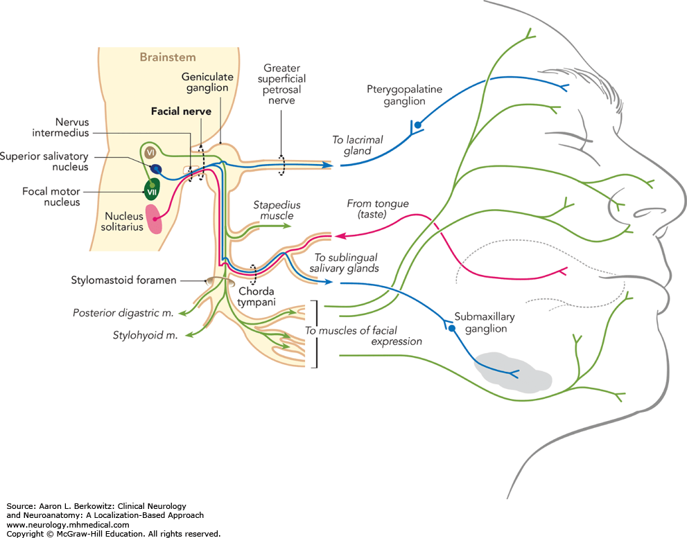
Cranial Nerve #7
The facial nerve(CN #7) sends taste sensations from the anterior part of the tongue to the brain. The motor pathway stimulates the muscles of the face and scalp. Bell’s palsy is a condition that affects CN #7 by causing muscle paralysis of one side of the face. The cause of Bell’s palsy is unknown, but it usually follows infections, such as Herpes, Measles, and Lyme Disease.

Cranial Nerve #8
The vestibulocochlear nerve (CN #8) consists of sensory neurons. The vestibular apparatus(a sense organ in your inner ear) only contains sensory neurons. The vestibular sense orients the position of our head to the body, which helps us maintain balance. The cochlear branch sends auditory information from the cochlea to the brain.

Cranial Nerve #9
The muscles involved in swallowing and saliva secretion receive stimulation via the glossopharyngeal nerve (CN #9) motor neurons. Tongue and throat sensations get to the brain via CN #9 sensory neurons. Also, taste sensations from the posterior part of the tongue travel to the brain via CN #9.

Cranial Nerve #10
The vagus nerve (CN #10) communicates with many major organs in the thoracic and abdominal cavities. The primary sensations are visceral, which are unconscious sensations of the internal organs. Visceral sensations from the eardrum, voice box (larynx), trachea, lungs, heart, esophagus, and the digestive tract travel to the brain via the vagus nerve. Vagus motor neurons stimulate skeletal muscles in the throat and larynx, cardiac muscle, and the smooth muscle surrounding the GI tract. CN #10 is the central nerve of the PyNS. Spinal nerves innervate the organs of the SyNS.

Cranial Nerve #11
Motor neurons in the accessory nerve (CN #11) stimulate the sternocleidomastoid and trapezius neck muscles. Patients with accessory nerve damage find it challenging to move their necks and raise their shoulders.

Cranial Nerve #12
The hypoglossal nerve (CN#12) comprises motor neurons that control tongue movements. Patients with hypoglossal nerve damage will display a tilted tongue. The misshaped tongue affects speech, swallowing, and breathing.

Summary
- The peripheral nervous system (PNS) comprises all of the neurons outside of the brain and spinal cord.
- The somatic nervous system is the only voluntary division of the PNS, and it controls skeletal muscle movements. Reflexes are a part of the somatic NS and are involuntary movements of skeletal muscles.
- The autonomic nervous system is involuntary and comprises the sympathetic NS and the parasympathetic NS.
- The sympathetic NS controls the expenditure of energy – i.e., fight or flight.
- The parasympathetic NS controls the conservation of energy – i.e., digestion.
- The spinal nerves begin at the spinal cord and extend to all regions of the body.
- There are 12 crainal nerves.
- Cranial nerves 1, 2, and 8 comprise sensory neurons.
- Cranial nerves 3, 4, 6, 11, and 12 comprise motor neurons.
- Cranial nerves 5, 7, 9, 10 comprise motor neurons and sensory neurons.