Your body comprises about 7 x 1027 atoms. Hydrogen, oxygen, carbon, nitrogen, and phosphorus make up 99% of all the body elements. Trace elements such as calcium, sodium, and potassium make up the remaining 1%. These elements are the foundation of all the organic and inorganic material that makes us human.
A Few Facts About Body Fluids
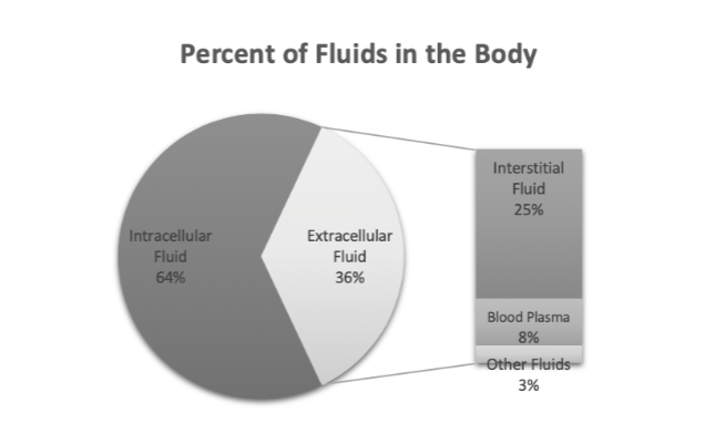
Water makes up about 65% of the total mass of your body. The solution found in your cells is known as intracellular fluid. Extracellular fluid is the fluid found outside of your cells, such as interstitial fluid (the fluid surrounding cells).
A substance or a solution that has a job is a secretion. For example, the secretion of hydrochloric acid helps the stomach enzymes digest protein. Excretion is a waste product. For example, your kidneys filter blood plasma to form the excretion of urine. Sometimes, a substance or solution can be secretion and excretion. For instance, sweat is a secretion because it cools us down when our body temperature rises above homeostasis (stability). Sweat contains waste products, too, so it is also an excretion.

Random Fact You Cannot Unlearn:
By your 21st birthday, you will have excreted 6,552 pounds of poop. That’s the mass of two beluga whales! Isn’t learning magical?

General Histology of Epithelial Tissues
Histology is the study of tissues. A tissue is when two or more different cell types interact. The four primary tissue types are epithelial, connective, muscle, and nervous. All body organs comprise two or more of these tissue types.
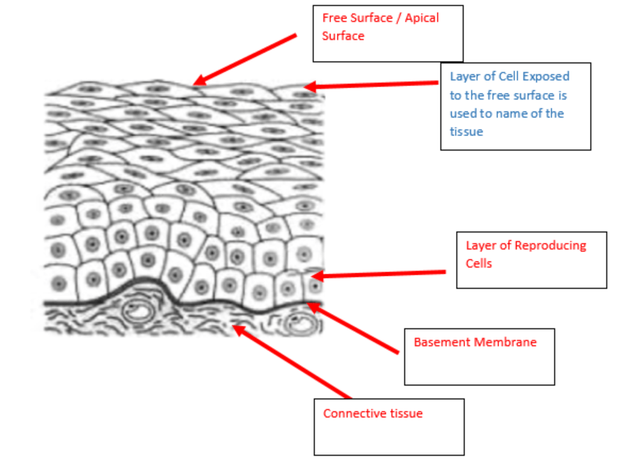
Epithelial tissues cover and line all external and internal body surfaces. Epithelial tissues consist of a basement membrane and a free surface. A basement membrane anchors the deepest layer of epithelial cells to connective tissue. An open space called the free surface or apical surface is present on the superficial side. Epithelial tissues contain few nerves and no blood vessels (avascular). So, the cell layer attached to the basement membrane is the only layer that receives enough nutrients for reproduction (cell division).
Epithelial cells regenerate rapidly because of the exposure to their free surface. Your skin is in near-constant contact with the world’s elements and needs a protective layer. The epidermis comprises a multilayered epithelial tissue with a superficial wall of death. The wall of death is not an extreme mosh pit but a layer of dead cells filled with a protective protein. The epidermis’ fast reproductive rate is why our skin does not bleed or become damaged when we put on a shirt or lay on the ground.
Lifespans of a Few Human Body Cell Types
| Cell Type | Average Lifespan | Cell Division |
|---|---|---|
| Stomach epithelium | 2 days | Yes |
| Small intestine epithelium | 1-2 days | Yes |
| Alveoli epithelium | 9 days | Yes |
| Skin epithelium | 10-30 days | Yes |
| Leukocytes (WBCs) | 12 hours – decades | Some types |
| Erythrocytes (RBCs) | 4 months | No |
| Fat cells | 8 years | Yes |
| Cardiac muscle | Lifetime | No |
| Skeletal muscle | Lifetime | No |
| Neurons | Lifetime | No (most types) |
Classifying Epithelial Tissue
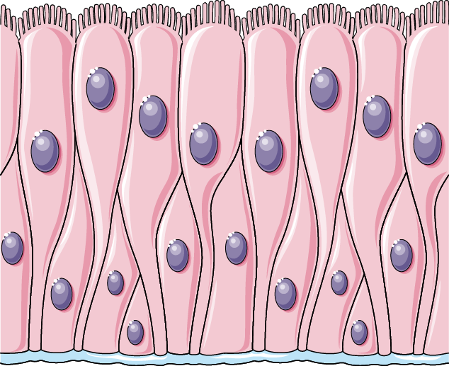
Epithelial tissues come in different layers. Simple epithelium refers to a single layer of cells that connect to the basement membrane and touch the free surface. Stratified refers to tissue with layers. Stratified epithelium comprises a deep cell layer attached to the basement membrane, followed by multiple cell layers that extend to the free surface. Pseudostratified epithelium appears stratified but only comprises a single cell layer – all cells connect to the basement membrane, but not all cells reach the free surface.
The Three General Types of Epithelium
| Tissue Type | Number of Cell Layers | Picture |
|---|---|---|
| Simple epithelium | One | 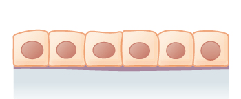 |
| Stratified epithelium | Two or more | 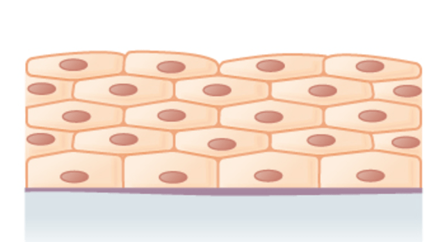 |
| Pseudostratified epithelium | One | 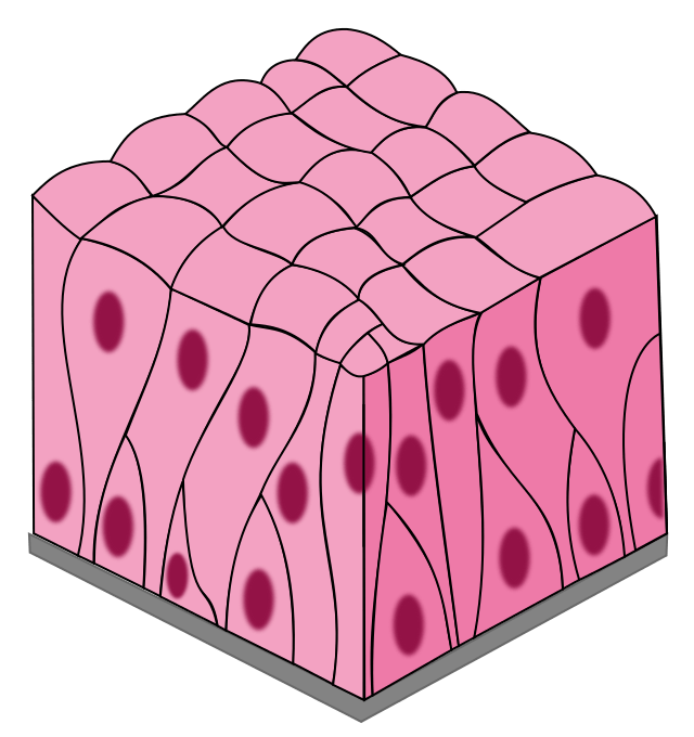 |

The pseudostratified epithelium is analogous to a forest. All the trees anchor to the ground (basement membrane) in a forest, but only some reach the canopy (free surface).
The Three Primary Cell Shapes
| Cell Type | Cell Shape | Picture |
|---|---|---|
| Squamous cell | Flat, thin |  |
| Cuboidal cell | Cube-shaped |  |
| Columnar cell | Column-shape |  |
Each layer of cells in the stratified epithelium is of a different shape. So, to classify stratified epithelium, you need to look at the shape of the superficial layer of cells (cells exposed to the free surface). Pictured below is an example of stratified epithelium. Notice how the cells attached to the basement membrane are cube-shaped. As the cells move towards the free surface, they become flattened. By the time the cells reach the free surface, they are flat. Therefore, the name of this tissue is stratified (has more than one cell layer), squamous (superficial layer of cells are flat), epithelium (lines the mouth).
Specific Epithelial Tissues
There are seven types of epithelial tissues that you have to know. Gas exchange, absorption, or secretion are the functions of the simple epithelium; secretion and the movement of substances are the functions of the pseudostratified epithelium; protection is the stratified epithelium function.
Gas Exchange Epithelial Tissue
The walls of the capillaries and the alveoli (tiny air sacs in the lungs) comprise a single layer of simple squamous epithelium. The thin walls allow for rapid oxygen and carbon dioxide exchange during respiration.


Picture Source: https://commons.wikimedia.org/w/index.php?search=simple+squamous&title=Special:MediaSearch&go=Go&type=image
Absorption and Secretion Epithelial Tissues
The walls of the tiny filters in kidneys, called nephrons, comprise simple cuboidal epithelium. This single layer of cube-shaped cells allows the absorption and secretion of water and solutes.


Picture Source: https://commons.wikimedia.org/w/index.php?search=simple+cuboidal&title=Special:MediaSearch&go=Go&type=image
Simple columnar epithelium lines the walls of the stomach, small intestine, and large intestine. In the small intestine, most food monomers move into the surrounding capillaries. Goblet cells secrete mucus, and their location is in between the columnar cells. The primary role of the mucus is to protect the epithelial tissue from acid and other chemicals.


Picture Source: https://commons.wikimedia.org/w/index.php?search=simple+columnar&title=Special:MediaSearch&go=Go&type=image
Secretion and Movement Epithelial Tissue
Pseudostratified columnar epithelium lines your trachea (windpipe) and bronchial tubes (airways in the lungs). Cells that extend to the free surface have small hair-like structures called cilia. Goblet cells secrete mucus that traps dust, smoke, and pathogens that we breathe in, and the cilia move these impurities up into the throat, where we swallow, cough, or sneeze out of the body.


Picture Source: https://commons.wikimedia.org/w/index.php?search=pseudostratified+columnar+&title=Special:MediaSearch&go=Go&type=image
Protective Epithelial Tissues
The superficial layer of skin is the epidermis, and it comprises keratinized stratified squamous epithelium (try saying this fast five times). The epidermis protects us from pathogens and ultraviolet (UV) radiation and prevents us from dehydrating. The superficial layer of cells is dead and packed with keratin. Keratin is the protein that gives the epidermis its protective and water-conserving properties.


Nonkeratinized stratified squamous epithelium protects the walls of the mouth, esophagus(the tube that takes food to your stomach), rectum, and vagina. This moist tissue does not contain keratin or a layer of dead cells.


Transitional epithelium is stratified, and it lines and protects the urinary bladder. Transitional epithelium can stretch, which changes the shape of the cells. When the bladder is empty, all the cells are cuboidal. As the bladder fills, the walls stretch, and the superficial layers of cuboidal cells transition into squamous cells.

Picture Source: https://i.pinimg.com/originals/71/e9/0e/71e90e311eca1663141e1034ccc1c8a7.jpg and https://commons.wikimedia.org/wiki/File:Anatomy_and_physiology_of_animals_transitional_epithelium.jpg
Key Concepts
- Epithelial tissues are superficial tissues that attach to a connective tissue via a basement membrane
- Epithelial tissues are avascular
- Epithelial tissues regenerate fast
- Simple epithelial tissues comprise a single cell layer
- Stratified epithelial tissues comprise multiple cell layers
- Pseudostratified epithelial tissues comprise a single cell layer but look stratified due to the different sizes and shapes of the cells
- Squamous cells are flat
- Cuboidal cells are cube-shaped
- Columnar cells are column-shaped
Great Video On Epithelial Tissues
The video below covers six out of the seven epithelial tissues you need to learn about (nonkeratinized stratified squamous). The jejunum is a part of the small intestine.