General Connective Tissue Histology
Connective tissue is the most abundant and diverse group of tissues in the human body. Because of connective tissues’ diversity, their anatomy and physiology depend on their location. For example, bone supports muscles and protects internal organs, blood transports gases and nutrients, adipose tissue insulates and stores energy, and dense fibrous connective tissue connects muscle to bones (tendons) or bones to bones (ligaments).
The Extracellular Matrix
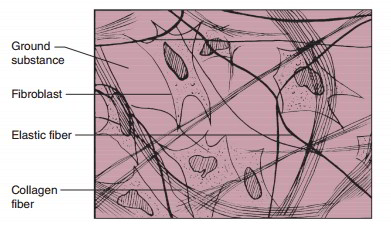
The two primary components of connective tissues are cells and the extracellular matrix. The extracellular matrix is a mixture of protein fibers in a clear, viscous liquid called the ground substance. The viscosity and composition of the ground substance vary between connective tissue types.

Protein Fibers
Protein fibers are in the extracellular matrix of all connective tissues except blood plasma. Collagen is the most abundant protein fiber in the body, accounting for 30% of protein. Collagen is a durable, bendable structural protein that gives connective tissue tensile strength. For example, your tendons are a sheet of collagen fibers that connect muscles to bones. Tendons must be durable to allow the muscles to move bones, sometimes against high opposing forces (lifting weights).
Elastic fibers give connective tissue elasticity. For example, the cartilage that makes up your outer ear (the pinna) has a high concentration of elastic fibers. The high concentration of elastic fibers is why bending the pinna will not affect its shape.
Reticular fibers comprise specialized collagen fibers that form the structural part of soft-tissue internal organs, such as the liver, spleen, and lymph nodes.
Cell Types in Connective Tissues
The cells within connective tissues vary, but fibroblasts are the most common cells because they make collagen and the bulk of the extracellular matrix. Erythrocytes (red blood cells) are the most abundant cells in the body, accounting for 70% of cells. However, erythrocytes are only in blood, whereas fibroblasts or fibroblast-like cells are in every other connective tissue.
Varying Degrees of Vascularity
Most physiology resources divide connective tissues into five categories: embryonic, connective tissue proper, bone, blood, and cartilage. But, we will classify the tissues by their vascularity, i.e., the degree of blood vessels in the tissue. Vascularity varies between connective tissues by being highly vascular (bone), slightly vascular (tendons), or avascular (cartilage).
Vascular Connective Tissues
Mesenchyme
Connective tissues are diverse but arise from the same embryonic tissue called mesenchyme, sans blood. Mesenchyme comprises mesenchymal cells(stem cell-like cells)that can differentiate into any connective tissue and an extracellular matrix with a few protein fibers. Mesenchyme is most abundant during embryonic development, is scarce in adults, and is mostly in the yellow bone marrow.


Areolar (Loose) Connective Tissue
Areolar connective tissue is a loose connective tissue because of the scattering of protein fibers throughout the ground substance. (I think it looks like the Jackson Pollock painting Summertime: Number 9A.) Fibroblasts are the most abundant cell, but leukocytes (white blood cells) and adipocytes (fat cells) are visible. The three protein fibers are present; collagen fibers are the amplest and most visible. Areolar connective tissue is beneath the skin and surrounding blood vessels, muscles, and nerves. Its primary function is to loosely bind organs together, such as wrapping nerves, blood vessels, and muscles together.


Adipose Tissue
Adipose tissue is fat. Here is what it looks like under the microscope:


There are two types of adipose tissue: (1) highly vascular brown adipose tissue (BAT) that can rapidly catabolize (break it down) calories into heat, and (2) white adipose tissue (WAT) that is not as vascular as BAT. WAT stores calories and is tough to catabolize. Newborns have more BAT than adults because they have less musculature and cannot shiver. When a baby gets cold, the rapid catabolism of BAT produces heat. Children and adults have better musculature and can shiver, so they have WAT and little BAT. Those who live in colder climates have more BAT, and those who can easily lose weight may have more BAT than those who have trouble losing weight.

Adipose tissue comprises adipocytes filled with fat droplets and a few scattered collagen fibers. The number of adipocytes remains constant throughout life. For every fat cell that dies, a new one takes its place.
If the number of fat cells remains constant, how do we gain and lose weight?
Your adipocytes gain and lose fat, which increases or decreases their volume. So, if you gain five pounds, you still have the same number of adipocytes, but they contain more fat.

The hypodermis is a layer of adipose tissue known as subcutaneous fat. The primary function of subcutaneous fats is insulation. During the winter months, our subcutaneous fat layer thickens to retain heat. The hypodermis thins during the summer months so we can release heat.

Visceral fat is the adipose tissue found within the abdominal and pelvic cavities. Small amounts of visceral fat surround and protect your internal organs (e.g., kidneys). There is a strong link between visceral fat percentage and metabolic disorders, such as type 2 diabetes. Individuals with less visceral fat levels are less likely to get a metabolic disease than those who are not overweight but have more visceral fat. The size of one’s belly can be a good indicator of overall health.

Adipose tissue’s principal functions are long-term energy storage, insulating the body, and protecting internal, soft-body internal organs – e.g., the kidneys and spleen.
Reticular Connective Tissue
Reticular connective tissue has a high concentration of leukocytes and reticular cells. Reticular cells are fibroblasts that make thick pieces of collagen called reticular fibers. The reticular fibers form a pattern that acts as a supportive skeleton for soft-bodied internal organs, such as the lymph nodes and liver.


Reticular connective tissue also comprises red bone marrow. The red bone marrow location is in the vertebrae and flat bones (ribs, ilium, skull bones) and the ends of long bones (femur, humerus). All bone marrow is red until the age of seven, and then half of the red bone marrow becomes yellow bone marrow over the next decade.

Hemopoiesis is the process of blood formation. During embryonic development, in the first two to eight weeks of pregnancy, hemopoiesis begins in the yolk sack. The liver becomes the primary blood-producing organ by the eighth week of gestation. At week 32, the red bone marrow becomes the blood-producing organ and remains so for life.
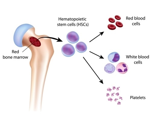
Red bone marrow contains hemopoietic stem cells that differentiate into 500 billion mature blood cells daily. Most of the newly made cells are red blood cells, with the remainder being white blood cells and megakaryocytes (cells that make platelets).
Bone
Bone is vascular because the blood is made in red bone marrow leaves via a blood vessel (bone marrow is not the same tissue as bone). Bone’s extracellular matrix comprises calcium, phosphorus, and collagen fibers. This mixture of organic and inorganic components gives bone its strength and flexibility. Gram for gram bone is stronger than steel and concrete. For example, it takes 4,000 newtons (N) or 900 pounds of force to break the femur but only 1,900 N to break concrete of the same mass. Well-trained martial artists can break 1.5-inch concrete slabs with their fists at 3,000 N—enough force to split the slab but not enough power to break a bone.
Random Boxing Fact
Former heavyweight boxer Frank Bruno could land a punch with a force of 6,300 newtons (1,400 pounds). This is enough force to speed up your head 53 times the force of gravity (53 g). The fictional boxer Ivan Drago from Rocky IV has a punch force of 35,580 N (8,000 pounds). His punch will accelerate a skull 303 times the force of gravity (303 g)–the same force as being hit by a 1975 AMC Pacer traveling at 30 mph.
Osteocytes, osteoblasts, and osteoclasts are the primary cells found in bone. Osteoblasts build the bone matrix, osteoclasts break down the bone matrix, and osteocytes manage the bone matrix. Osteocytes live in small holes called lacunae, interacting with one another through tiny canals called canaliculi.


Bone stores 99% of the body’s calcium. The stored calcium regulates our blood calcium levels to prevent hypocalcemia and hypercalcemia. When we are hypocalcemic, osteoclasts become active and break down the bone matrix, which releases calcium into the blood. When we are hypercalcemic, osteoblasts use the excess calcium to build bone. Bone protects most of our major internal organs. For example, the bones of the skull protect your brain. Bones are the primary support structures of our bodies, too. Without bones, we would be a pile of soft tissue.

Blood
Blood ( is in blood vessels, so by default, it is vascular. Blood comprises two main parts: formed elements and plasma. The formed elements are erythrocytes, leukocytes, and platelets. Erythrocytes comprise 45% of the total blood volume, and their function is gas transport. Leukocytes comprise less than 1% of the blood volume because they are primarily in other tissues, such as reticular connective tissue. Leukocytes are the primary cells of the immune system. Platelets(thrombocytes) are fragments of a former cell that comprise less than one percent of the total blood volume. Thrombocytes are critical in hemostasis (the stoppage of bleeding). Plasma is the extracellular matrix of blood and makes up the remaining 55% of blood volume. Plasma is 92% water with many solutes (gases, ions, glucose, amino acids, fatty acids, steroids, non-steroid hormones, etc.), but blood lacks protein fibers.

Slightly Vascular Connective Tissues
Dense Regular Connective Tissue
Dense regular connective tissue is a sheet of collagen fibers arranged in parallel to one another with a few fibroblasts scattered throughout it. This collagen arrangement allows this tissue to withstand high tensile force in a single direction. For example, dense regular connective tissue forms the tendons and ligaments of your body. The high tensile force of tendons and ligaments plays a significant role in moving the skeleton.


Dense Irregular Connective Tissue
The dermis is the thickest layer of skin and is deep to the epidermis. The dermis comprises dense irregular connective tissue.


Dense irregular connective tissue has many collagen fibers that are not parallel to each other but arranged in an irregular pattern that resists tearing. This irregular arrangement of collagen fibers is why it takes a lot of force to rip your skin. But, age and prolonged exposure to ultraviolet radiation will weaken the dermis, making it easier to tear.


Pictured to the left is your Physiology teacher at age 18. Pictured to the right is a current picture of your Physiology teacher. Please use sunscreen, or this is what you may look like when you are 49.
Avascular Connective Tissues
Cartilage is avascular. Since cartilage does not have a direct blood supply, it takes a long time to heal when damaged. Cartilage has cells called chondrocytes inside lacunae. Chondrocytes are like fibroblasts because both make protein fibers found in the extracellular matrix.
Hyaline Cartilage
Hyaline cartilage is the most abundant cartilage. It makes up the distal part of your nose, the cartilage connecting your ribs to your sternum (costal cartilage), the C-rings that support your trachea (windpipe) and bronchial tubes (tubes in lungs), most of the fetal skeleton, growth plates in bones, and the ends of bones at moveable joints. Hyaline cartilage has a shiny matrix made of fine collagen fibers that create a smooth surface. Hyaline’s smoothness provides an ideal surface for two bones to move across each other’s surface with little effort.


Elastic Cartilage
Elastic cartilage supports the outer ear (pinna). Elastic cartilage has collagen fibers and lots of elastic fibers, giving the pinna high elasticity.


Fibrocartilage
The extracellular matrix of fibrocartilage comprises a thick mat of collagen fibers with chondrocytes in lacunae. Fibrocartilage functions as a shock-absorbing and compression-resistant tissue, adding these properties to the knee joint (meniscus) and the vertebral column (intervertebral discs).


Key Concepts
- Connective tissues have a degree of vascularity: vascular, slightly vascular, or avascular
- All connective tissue comes from mesenchyme, sans blood (blood comes from hemopoietic stem cells)
- Connective tissues comprise the non-living extracellular matrix and cells
- Connective tissues are the most diverse and abundant group of tissues