Calcium is More Than a Pet Turkey on a Hat
So, are you ready to learn more about membrane potential?

Is that a yes?

It includes calcium.

Calcium!!!!!!

This is a picture of my pet turkey, Calcium, on a hat.

No. . . What?! There is something seriously wrong with your neural networks.
Could be calcium.
You do know you are arguing with yourself, right?
I’m Tyler Durden.
No, you are not.
I’m Batman?
No.
Gobble gobble?
That’s more like it.
Section 1: What’s the Big Deal with Calcium?
So far, you have learned that the bones store calcium and the parathyroid gland, and, to a lesser extent, the thyroid gland regulates the levels of calcium in the blood plasma (the liquid part of the blood). Calcium also plays a critical role in releasing neurotransmitters from the terminal ends of neurons. In cardiac muscle, calcium ions are essential for:
- It makes the union of the contractile proteins possible
- Prolongs depolarization by 200-fold so the heart can contract
Section II: The Cardiac Conduction System
There is a scene in Indiana Jones and the Temple of Doom where the main antagonist rips a person’s heart out of their chest with his hand. The victim stares in horror as his heart beats in the antagonist’s hands. The heart continues to beat until it catches on fire. Now, a human can’t use their bare hand to rip a heart out of a human’s thoracic cavity. And hearts usually do not spontaneously catch on fire. However, there is a little nugget of truth to this scene. If given a constant oxygen-rich blood supply, the heart can set its rhythm and hypothetically maintain a heartbeat outside the body.

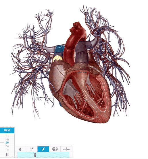
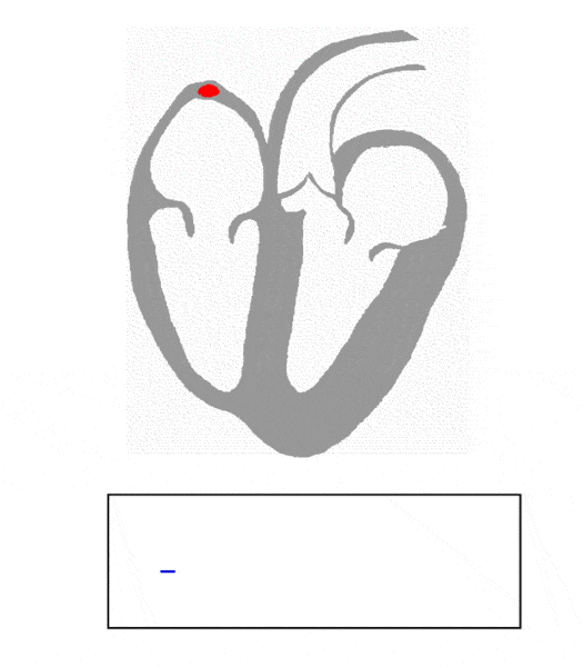
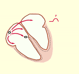
The cardiac conduction system comprises specialized cardiac fibers that send action potentials through the myocardium, causing the heart to contract. In the upper wall of the right atrium sits a cluster of specialized self-exciting cardiac fibers known as the sinoatrial node, or SA node. The SA node is the heart’s pacemaker, setting the heart’s rhythm at about one heartbeat per second. The action potential initiated by the SA node travels through the atrial walls, stimulating the myocardium’s contraction. The action potential arrives at another bundle of self-exciting cells called the atrioventricular node or AV node. At the AV node, the action potential pauses so that the contracting atria can pump the remaining blood into the ventricles. The AV node releases the action potential, and it travels to the myocardium in the interventricular septum via the bundle of His. The bundle of His splits into the left and right bundle branches, and the action potential travels down both pathways. The right bundle branch leads to the Purkinje fibers in the walls of the right ventricle, and the left branch leads to the Purkinje fibers in the walls of the left ventricle. The action potential travels along the Purkinje fibers, stimulating the ventricular myocardium—the ventricles contract, pumping the blood into the arteries. The heart relaxes, and the process repeats when the SA node spawns a new action potential.
Here are three GIFs of the cardiac conduction system:


The medulla oblongata stimulates the SA node via the neurons of the autonomic nervous system. However, the medulla only controls heart rate, not heart rhythm. A decrease in heart rate results from the neurotransmitter acetylcholine released from neurons in the parasympathetic nervous system. An increase in heart rate results from the neurotransmitter norepinephrine released from neurons in the sympathetic nervous system.

Your heart contracts and relaxes as a unit because of cardiac fibers’ connection by specialized gap junctions called intercalated discs. The intercalated discs allow the cardiac action potential to flow from the top of the right atrium to the heart’s apex (bottom of the left ventricle).

Section III: Calcium’s Effect on the Cardiac Membrane Potential
The cardiac muscle cell membrane potential is similar to the neuron membrane potential. However, key differences exist between the membrane potentials of cardiac muscle cells and neurons. The primary difference is the role of calcium ions (Ca2+) in cardiac membrane potentials.
Cardiac Muscle Fiber Resting Potential
The cardiac muscle resting potential shares the following properties with the neuron resting potential:
- The resting membrane potential is a negative charge since there are more anions than cations inside a cardiac cell (aka cardiac fiber)
- Sodium (Na+) concentration is highest outside a cardiac cell, and potassium (K+) concentration is highest inside a cardiac cell
- Ca2+ concentration is highest outside the cardiac fiber
- The equilibrium potential is a product of the chemical gradient and the voltage gradient
- The Nernst equation calculates the equilibrium potential of each ion species
The cardiac resting membrane potential differs from the neuron resting potential via the following property:
- A higher concentration of Ca2+ is inside the cardiac fiber in an organelle called the sarcoplasmic reticulum
The Ca2+ extracellular concentration refers to the Ca2+ outside of the cell and the Ca2+ inside the sarcoplasmic reticulum. The sarcoplasmic reticulum is in the intracellular fluid, but the Ca2+ it contains does not affect intracellular Ca2+. Since a membrane surrounds the sarcoplasmic reticulum, its Ca2+ has the same effect as the extracellular fluid. Therefore, the Ca2+ extracellular concentration combines the Ca2+ outside the cell and inside the sarcoplasmic reticulum.

When a cardiac fiber is at rest, no Na+ voltage-gated channel proteins (VGCPs) and Ca2+ VGCPs proteins are open, and a few K+ channel proteins are open.
Cardiac fiber action potential terminology is similar to neuron terminology, but there are a few key differences. Below is a table of similarities and differences between action potentials in neurons, the conductive cardiac fibers (fibers that make up the SA node and AV node), and the contractile cardiac fibers (fibers of the myocardium).

Cardiac CONTRACTION Fiber Action Potentials
The myocardium that surrounds the chamber of the heart comprises cardiac contraction fibers. Cardiac contraction fiber action potentials differ from the action potentials in the SA and AV nodes. The resting membrane potential is stable like neurons, requiring stimulation from the SA node or AV node to reach a threshold.
The action potentials in the cardiac fibers that surround the atria and ventricles occur in four stages:
- Depolarization to the threshold: A cardiac fiber has a threshold potential like a neuron. However, unlike a neuron, the threshold potential is not the product of the summation of graded potentials (IPSPs and EPSPs). Instead, the action potential from the SA node and AV node stimulates the cardiac fibers. During this phase, Na+ VGCPs open, and Na+ trickles into the cell until the threshold. At the threshold, all Na+ VFCPs open, and rapid depolarization begins.
- Rapid Depolarization: Na+ VGCPs open and close rapidly. The influx of Na+ reverses the membrane potential, and a cardiac cell’s inside becomes positive, and the outside becomes negative. Rapid polarization is so fast that the whole stage happens before the heart’s contraction.
- Prolonged Depolarization (The Plateau Stage): Extends depolarization in a cardiac contractile fiber and allows for the heart’s complete contraction. Prolonged depolarization starts when Ca2+ VGCPs open in the cell membrane and the membrane of the sarcoplasmic reticulum. Ca2+ extends depolarization from one millisecond to 200 milliseconds. Prolonged depolarization is not static. A slow drop in voltage happens during the partial diffusion of K+ out of the cell and the sodium-potassium pump removing Na+ from the intracellular fluid. However, the flow of Ca2+ keeps the membrane potential depolarized enough to form a plateau on an action potential graph and allows for the complete contraction of the heart. Once the Ca2+ VGCPs close, all the K+ VGCPs open, and repolarization begins.
- Repolarization: The heart begins to relax with repolarization. During repolarization, all K+ VGCPs open, and K+ leaves the cell. The inside of the cardiac contractile fibers becomes negative, and the outside becomes positive. The sodium-potassium pump and calcium pump return the ions to their resting state, and the cell returns to resting potential.


Cardiac Fibers Have a Long Refractory Period
Cardiac contractile fibers have an absolute refractory period that allows the ventricle to eject most of the end-diastolic volume, preventing the heart from entering tetanus. Tetanus is a sustained muscle contraction. Skeletal muscles achieve tetanus all the time due to their short refractory period, and this is how our core muscle keeps us upright. However, if cardiac fibers were to become tetanic, the heart would stop pumping blood.


Section IV: Linking the ECG to Heart Physiology
A trained physician, nurse, or medic can learn a lot about a patient’s heart function by looking at an ECG trace. This section will examine one cardiac cycle on an ECG trace.
An atrial action potential occurs from the start of the P wave and ends at the S wave. The P wave denotes atrial depolarization, and the QRS complex represents atrial repolarization. The PR interval begins at the start of the P wave and ends at the beginning of the QRS complex, and it is during this interval the atria contract. The RP interval implies atrial diastole, which begins at the R wave and ends at the top of the P wave of the next cardiac cycle.
The P wave shows the health of the SA node. A functioning SA node will produce an arched P wave with a duration of 0.06-0.12 seconds and a voltage of 0.1-0.2 mV.
The QT interval on an ECG trace represents a ventricular action potential. The QRS complex represents the ventricles’ rapid depolarization. The ST segment shows prolonged depolarization (plateau stage), and the T wave denotes ventricular repolarization. Ventricular systole begins at the S wave and ends after the T wave. Ventricular diastole occurs during the TR interval, right after the T wave to the R wave in the next cardiac cycle.
The Q-T interval is present when the AV node is functioning. Irregularities with the QT interval indicate problems with ventricular systole.
ECG Data Table

Click here for an excellent reference on ECG basics and more advanced ECG information.
How to Read an ECG


Summary
- An ECG trace is an accumulation of cardiac action potentials showing events during cardiac cycles.
- The sinoatrial (SA) node sets the rhythm of the heart = happens at the start of the P wave
- The atrioventricular (AV) node delays the action potential, so the atria contract and then release it to initiate ventricular systole = PR segment
- P wave = Atrial depolarization
- PR interval = Atrial systole
- QRS complex = Rapid ventricular depolarization via the influx of Na+
- ST segment = Prolonged depolarization (plateau stage) via the influx of Ca2+
- T wave = Ventricular repolarization via the efflux of K+
- QT interval = Ventricular systole
- RP interval = Atrial diastole
- TR interval = Ventricular diastole