Cardiac muscle fibers have the contractile proteins actin and myosin arranged in units called sarcomeres. Sarcomeres are the contractile units of cardiac fibers. Each muscle fiber contains many sarcomeres arranged in cylindrical myofibrils. A web-like structure called the sarcoplasmic reticulum surrounds the myofibrils and releases Ca2+ during heart contraction.
Actin and myosin are parallel to each other and overlap one another. Actin comprises small, spherical protein monomers bound together, forming two long polymer chains. The two chains overlap each other and form a thin helix-shaped protein called the actin filament.
Myosin comprises many bound golf club-like monomers that form a thick, cylindrical polymer called a myosin filament. Myosin filaments have globular protrusions, or globular heads, that dot and surround the polymer. The globular heads are in pairs, which form a cross-bridge. Cross-bridges are where the myosin filament reacts with an actin filament.
Each spherical protein of an actin filament has a little notch on its superficial surface called the myosin-binding site. A cross-bridge on a myosin filament fits into the myosin-binding site when the cardiac muscle fiber is contracting. However, when a cardiac muscle fiber is relaxed, a protein called tropomyosin blocks the myosin-binding site. The only thing that can move tropomyosin is calcium. Without calcium, the actin and myosin filaments cannot form bonds, and all the sarcomeres within a muscle fiber will be unable to contract.
Here is how cardiac contraction works:
| 1. Cardiac fiber enters the plateau stage, causing Ca2+ to enter the intracellular fluid. | |
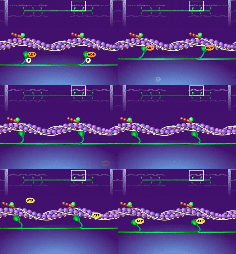
| 2. Ca2+ binds to tropomyosin, making the myosin-binding sites accessible. | ![Breakdown of ATP and Cross Bridge Movement during Muscle Contraction [HD Animation] animated gif](https://j.gifs.com/vVbXar.gif) |
| 3. The myosin cross-bridges bind to the myosin-binding sites and pull the actin filaments inward (power stroke) |  |
| 4. The inward movement of the actin filaments shortens the sarcomeres – i.e., muscle contraction. |  |
| 5. The cardiac fiber uses ATP to break the actin-myosin bond, which reloads the cross-bridges. |  |
| 6. Steps 3-5 repeat until the cardiac fiber contraction is complete, and relaxation begins. |  |
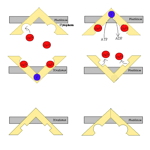
| 7. Ca2+ leaves the cardiac fiber via active transport (Ca2+ pumps) |  |
| 8. The absences of Ca2+ leads to tropomyosin blocking the myosin-binding sites – i.e., acting and myosin filaments can no longer form bonds. | |
| 9. All the sarcomeres return to their resting state, and the cardiac fiber relaxes. |
The nine steps above repeat with each cardiac cycle (heartbeat).
Calcium is critical for the proper function of the heart. During prolonged depolarization, the influx of Ca2+ extends the action potential by 200-300 milliseconds, which is the time required for the actin and myosin filaments to react with each other (steps 1-6 above).
A quick note about preload and contractility. The more cross-bridges that form between actin and myosin, the greater the preload and contractility of a cardiac fiber; therefore, the more actin/myosin power strokes there are, the higher the stroke volume and cardiac output.