A Random Stream of Consciousness
Whenever I hear the song “Smells Like Team Spirit” by Nirvana, my thoughts wander to the first time I heard it. The year was 1991, and I was a senior in high school. Yes, I am that old. I was in the passenger seat of my friend Tif’s car, and we were waiting for the stoplight to turn green. I remember the smell of incense, The Stone Roses cassette on the floor, and the scuff marks on my Doc’s resting on the dashboard. I feel almost the same level of anticipation that Tif and I felt as we waited for the Live 105 DJ to announce the song’s name and artist. These memories and feelings last the length of the song and fade with the song’s final chord. How is it possible that a single song can spark so many beautiful memories and feelings? How can one song transport me to a time when my hair was dirty blonde, my face was wrinkle-free, and my closet filled was with thrift-shop flannels? Why are the memories cathartic? Is it magic? Is it spiritual? Is it the flow of ions and chemicals of neural networks within the brain? Or is it a potpourri of the physical and metaphysical?

The Claustrum
The basic scientific definition of consciousness is being awake and aware of your surroundings. Why humans have it, and the origin of its existence is unknown. Duelists believe the conscious mind exists in a metaphysical plane, while hard determinists believe that it is an illusion produced by neural networks within the brain. Some believe it evolved to give us a purpose to survive and reproduce. Others state that there is a 50-50 chance we are living in a computer simulation. Some believe consciousness is a gift from the gods or that the answers we seek are in a mushroom-induced altered state.
Studies show that arousal (awake), the first step in consciousness, begins in the brain stem. However, the origin of awareness is much more difficult to locate. Recent research has focused on a small group of neurons called the claustrum as a possible origin of awareness. The claustrum’s neurons are unique because they make connections with all brain’s cortical regions (conscious centers). When scientists stimulate the neurons of the claustrum, patients lose awareness, but not arousal and become zombie-like. When the stimulation stops, the patients become fully aware and have no memory of the zombie-like event. Does this mean the claustrum is the origin of awareness? No. But, research may unlock the answer soon.
The 10% Factoid
A common factoid in our culture is that humans only use ten percent of their brains. The short answer to this is WRONG!!!!! Humans and all animals with a brain-like structure use the entirety of their brains. Each part is necessary for the brain to function as a whole. However, the ten percent factoid touches upon a mysterious phenomenon. This phenomenon is: why can some people do extraordinary cognitive tasks that most cannot? For example, let’s discuss Einstein’s brain. The assumption at his death was that Einstein used more of his brain than most humans. However, Einstein’s genius in mathematics may have been due to a loss of brain function. Einstein’s brain was missing a part of the frontal lobe that plays a role in language. The hypothesis is that his brain compensated for the loss by strengthening neural networks in the brain’s visuospatial region. Einstein’s brain’s visual-spatial area had a higher concentration of glial cells and neural connections than the average brain. However, neither the loss nor gain in brain anatomy proves that either of these regions was the origin of his genius. Einstein’s brain did not have more neurons or a greater mass than the average male of his size. His brain had a few abnormalities, but so do all brains. As the saying goes: what makes you different may help you establish the most famous scientific equation that very few people understand. I’m paraphrasing, of course.

The Divisions of the Brain
The brain makes up about two percent of our body mass but consumes 20 percent of blood oxygen and glucose when resting. The more complex the task, the more brain regions and energy are required to execute it. Most neurology textbooks divide the brain as follows:
- Telencephalon = The cerebrum
- Diencephalon = Portion that sits atop the brain stem
- Mesencephalon = Superior portion of the brain stem
- Metacephalon = Middle portion of the brain stem and cerebellum
- Myelencephalon = Inferior portion of the brain stem


Other sources use three regions to describe brain function:
- Cerebrum = The cognitive, conscious centers
- Cerebellum = Coordination and balance centers
- The Brain Stem = Vital and involuntary functions
However, this chapter will separate the brain into the following sections:
- Cerebrum = The cognitive, conscious centers
- Limbic system = The emotional and motivational centers
- Diencephalon = Behavioral centers and sensory filters
- Cerebellum = Balance, coordination, and posture centers (and probably consciousness, too)
- Brain stem = Vital function centers (heart rate, respiration, blood pressure)
The Cerebrum
Cognition and consciousness require complex and vast neural networks, so the cerebrum comprises eighty percent of the brain’s mass. The cerebrum consists of a thin outer layer of gray matter called the cerebral cortex and a dense web of myelinated axons. The cerebral cortex has an average thickness of 3 millimeters and a surface area of up to 200 square centimeters – that’s six square feet! Folding the cerebral cortex into peaks and valleys is how its large surface area fits into the skull’s confines. The sulci, or fissures, are the cerebral valleys, and the gyruses are the peaks. These gyri and sulci make up the five lobes of the cerebral cortex.
The Cerebral Hemispheres
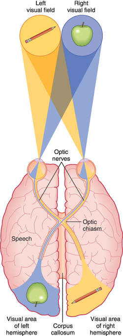
The cerebrum has a left hemisphere and a right hemisphere. The left hemisphere controls the right side of the body, and the right hemisphere controls the left. The two hemispheres communicate via a large bundle of myelinated axons called the corpus callosum.
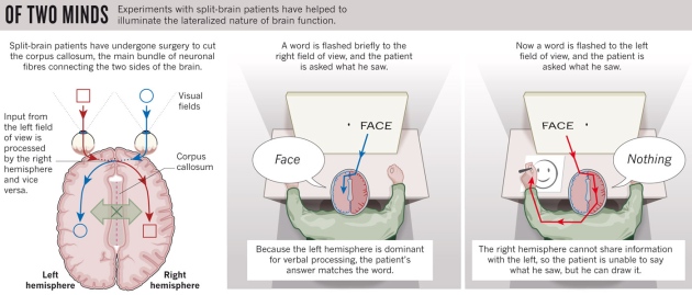
Before the availability of antiseizure medications, one of the best treatments for severe epilepsy was severing the corpus callosum. The severing of the corpus callosum stopped the seizures but also halted communication between the two cerebral hemispheres. People with epilepsy who had this procedure done to them are known as split-brain patients.
The most prominent symptoms of split-brain patients involve language. When split-brain patients see images in the right field of view, they can say what they are. However, they see nothing when the images are in the left field of view. (Remember, the left hemisphere controls the right side of the body, and the right cerebral hemisphere controls the left.)
Does this mean there is damage to their optic nerves or the brain’s visual centers?
Nope. The left hemisphere contains the language centers. When viewing an object in the right field, the left hemisphere can say its meaning. The right hemisphere is mute, so when it sees the item via the left field of view, it sends the information to the left hemisphere for processing. However, the right and left hemispheres cannot communicate without a corpus callosum. Without communication, a person cannot say what the object is in the left field of view or if an object is present. Now, this is where it gets weird. When split-brain patients grab a specific item in their left field of view, they grab the correct object even though it is not consciously perceived. Asking split-brain patients why they picked the object, they confabulate a reason why. Confabulating a story about the object proves that the right hemisphere can correctly process the visual information it receives. It just cannot verbalize it.
The Frontal Lobe
The frontal lobe is the largest cerebral lobe of the brain. Its functions include but are not limited to, conscious motor movements, problem-solving, judgment, language, memory, impulse control, and appropriate human behavior. The neurons’ product within the anterior portion of the frontal lobe is judgment, impulse control, and problem-solving. The emotional segment of the brain, the limbic system, is directly connected to the frontal lobe. However, the neurons in this network do not reach full myelination until people are in their mid-twenties to easily thirties (this is especially true for males).
Why?
Nerve impulses traveling lightly myelinated axons take longer to get to their destination. The underdeveloped myelin is why adolescents or young adults are more likely to act on their emotions than middle-aged adults. Unfortunately, traumatic brain injuries, such as abuse during childhood or multiple concussions, may prevent the full myelination of limbic-frontal connections. (The majority of violent felons have a traumatic brain injury, primarily frontal lobe damage.)

Primary Motor Cortex
The central sulcus is the longest sulcus/fissure in the coronal plane, separating the frontal and parietal lobes. The primary motor cortex (PMC) is the frontal lobe’s posterior gyrus, right before the central sulcus. Conscious skeletal muscle movements originate in the PMC. The PMC has an arrangement similar to a topographical map called a homunculus. Some parts of the body receive more PMC real estate than others. For example, hand and facial muscles have larger areas on the PMC due to the fine motor movements of hand and facial gestures. The back and chest muscles have smaller regions due to their gross motor movements.
The left motor cortex controls the right side of the body, and the right motor cortex controls the left side. Damage to this area can lead to paralysis of skeletal muscles. However, reflexes and involuntary muscle movements, such as breathing, may not be affected.

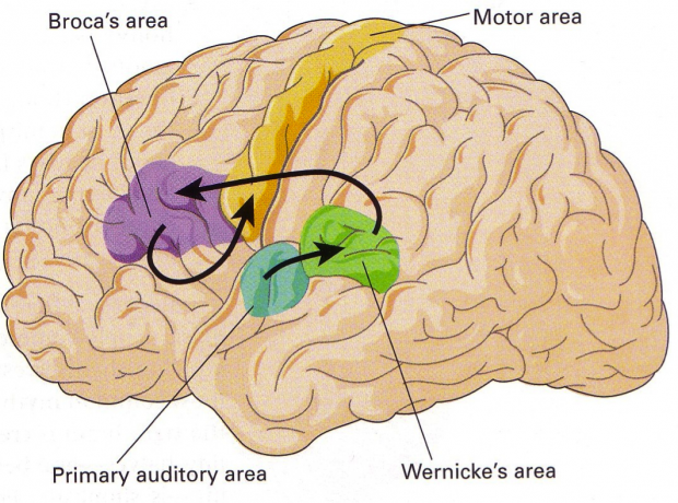
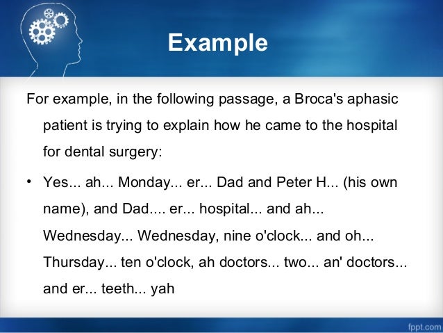
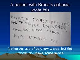
Language is almost exclusively a left hemisphere function. Anterior to the left posterior motor cortex is a small piece of cortex called Broca’s area. Broca’s area controls muscle movements needed for language expression. Patients with Broca’s aphasia have little to no loss in their mouth and throat muscles during mastication (chewing) and deglutition (swallowing). However, once they try to speak, their speech is effortful and primarily consists of nouns. All forms of language suffer, including sign language and written communication. Broca’s aphasia patients can use their hands to do all tasks except language. They may be able to tie their shoes, but they can barely communicate via writing or sign language.
So, they can use their hands to knit?
Yep.
What about typing on a keyboard?
If they are expressing language, no. If they are randomly hitting keys, yes. However, each patient’s symptoms will vary depending on the extent of the brain damage. Some patients can somewhat articulate via speech or written language, while others can only produce a single word or nonsense word.
The Temporal Lobe
Wernicke’s area is in the posterior region of the upper left temporal lobe. One of the temporal lobe’s primary functions is processing conscious auditory sensations. Wernicke’s area is responsible for comprehension in speech, which involves listening to sounds. Wernicke’s aphasia is a condition usually caused by a stroke. A patient’s speech is often incomprehensible and contains a mixture of real and made-up words (sometimes called word salad).
Wait, how is Wernicke’s aphasia different than Broca’s aphasia?
Below is a table that enumerates the differences between the two aphasias.
| Broca’s Aphasia | Wernicke’s Aphasia | |
|---|---|---|
| Speech articulation | No or partially | Yes |
| Speech comprehension | Yes, except written words need to be in nouns. For example, patients usually cannot read “to be or not to be” but can read “two bee oar knot two bee.” | No |
| Awareness of condition | Yes | No |
| Affects written and verbal communication | Yes | Yes |
The songs, voices, and noises you hear are the production of neural networks within the temporal lobe.
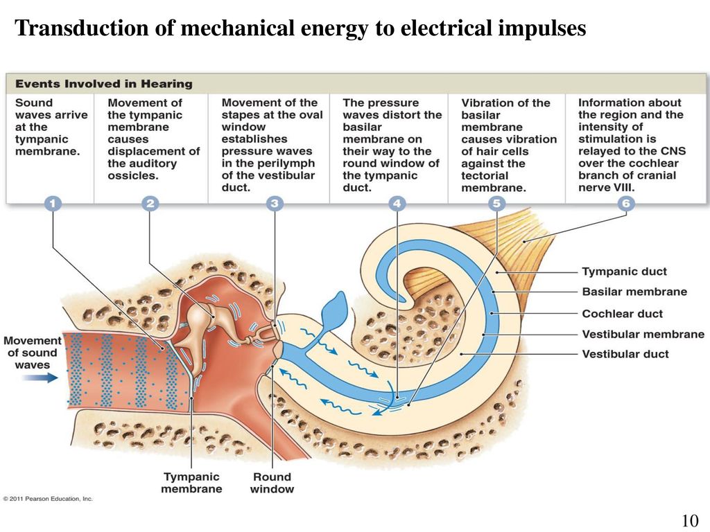
But how does the temporal lobe receive auditory sensations?
It begins when a person greets you with, “Hello.” The sound waves leave their mouth and travel to your ears. The sound waves vibrate the tympanic membrane (eardrum), which trembles the tiny bones in your ear (ossicles). The quivering ossicles push the liquid in the cochlea, and the motion of the fluid bends neuron-like hair cells. The hair cells’ bending excites neurons in the vestibulocochlear nerve (CN#8), sending the sensory information to the brain. The temporal lobe receives and processes the auditory sensations; you perceive the sound as the word hello.
The temporal lobe is located just behind the temples, hence its name. It is separated from the lateral cerebral sulcus’s frontal and parietal lobes, the largest sulcus in the transverse plane.
The Parietal Lobe
The parietal lobe is between the frontal and occipital lobes and is superior to the temporal lobe. The anterior gyrus sits directly after the central sulcus and is home to the somatic sensory cortex (SSC). The SSC is similar to the PMC in that some body areas receive more cortex than others. However, the SSC processes conscious tactile sensations, not motor movements.
Proprioception is your ability, both consciously and unconsciously, to know where your body is in space. The parietal lobe is responsible for the conscious part, so you can scratch the back of your head. Without proprioception, people cannot move a body part if it is not in their line of sight. For example, thank proprioception if you can walk without looking at your lower limbs and feet. People who lose proprioception can only walk when looking at their legs and feet. When their gaze wanders from their legs, the muscles go limp, and they fall.
Reading a map (visuospatial skills), understanding numbers, and knowing the location of your parked car without seeing it are other functions of the parietal lobe.

The Occipital Lobe
The posterior portion of the cerebral cortex is the primary visual cortex or occipital lobe.
The occipital lobe receives sensory information from the optic nerve. Vision is our most complex sense, so a whole lobe is responsible for processing visual data. Color, shape, movement, and spatial visual sensations are combined in the occipital lobe to form images.

A stroke in the visual cortex may lead to a condition called visual agnosia. Patients suffering from visual agnosia see images in fragments, not as whole parts. They can see an object but not recognize it, even their smartphone.
Wait. They cannot visually recognize their phone?
Usually.
How do they survive?
By eating, drinking, and breathing.
But how do they live without Instagram?
I survive without social media, and my vision works.
But you are a teacher. I asked, how do they live?
I live. I spend a lot of time gardening. Gardening is living, right?

Is that the emoji for the book The Man Who Mistook His Wife For a Hat?

Oh, I know this emoji! It’s the caninus, zygomaticus, mentalis, quadratus labii interioris, trangularis, buccinator, orbicularis muscles emoji. Or, as your generation calls it, the CZMQLITBO emoji. Putting phrases into acronyms makes it radical to the max.
How long is this conversation going to last?
It depends if we are still discussing gardening.
We never were.
Then, I guess we’re done. I bid you farewell with the universal goodbye emoji:
Please stick to gardening.

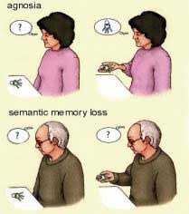
![Visual agnosia] You can see things. You copied the picture perfectly. Therefore, you do not have any problems with your eyes. Howev… | Told you so, Cognitive, Face](https://i.pinimg.com/originals/cd/bf/ea/cdbfeacdac3ff455b3086a79f21d387e.png)
The Insula
The insula, meaning island, is a deep layer of cortex found under the lateral cerebral sulcus. Studying the insula has been difficult due to its location. However, with modern brain imaging techniques, some of the functions of the insula are now known. The insula processes the conscious sensations of smell, taste, and balance. It allows us to perceive pain – pain is a perception, not a sensation – and our sense of self originates in the insula.


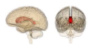
The Limbic System
The limbic system is responsible for motivational and emotional behaviors. The two main structures of the limbic system are the amygdala and hippocampus.

The amygdala is an almond-shaped cluster of cell bodies that process raw, unfiltered emotions. The primary emotions it regulates are fear and anger. Damage to this area can lead to a lack of fear or uncontrollable rage, aggression, or fear.
Our most vivid memories are emotional because there is a strong connection between the amygdala and the hippocampus. The hippocampus is critical for learning and memory and is one of the only brain areas that gain neurons throughout life. The hippocampus’s primary job is converting short-term memories into lasting long-term memories. Damage to the hippocampus can cause anterograde amnesia, the inability to form new long-term memories. People with severe bilateral hippocampal damage only have memories before the injury. Any new fact, person, or location will never become a lasting memory. The movie “Momento” (arguably Christopher Nolan’s best film) does an excellent job showing hippocampal damage, and the movie “50 First Dates” makes light of it. (However, this is a severe condition, and patients require constant care.)
This video is really detailed and mentions brain regions you do not have to know.
The Diencephalon
The hypothalamus, pineal gland, and thalamus are the three primary structures of the diencephalon. As you learned in Unit 3, the hypothalamus controls most of the endocrine system (hormones). Also, the autonomic nervous system is under hypothalamic control.
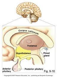
The pineal gland is the only major structure that is not bilateral. The singularity of the pineal gland is the reason why the French philosopher, Mathematician, and Frank Zappa impersonator, Rene Descartes hypothesized that it housed the human soul. We now know that the pineal gland does not contain the soul, but it helps the hypothalamus regulate the circadian rhythm (sleep-wake cycle) via the release of the hormone melatonin.
The thalamus is the cerebrum’s chief of staff. All sensory information, except olfaction, passes through the thalamus before reaching the cerebral cortex. The thalamus filters sensory information, so only the most crucial information makes it to our consciousness. Therefore, you will never perceive most of the sensory information your sense organs collect. Being unaware of most sensory stimuli may seem like a hindrance, but it is critical for learning and survival. Too many sensory stimuli can overload our consciousness, a state called sensory overload. Sensory overload is common among those living with autism.
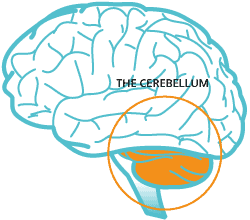
The Cerebellum
The cerebellum is a walnut-sized area of the brain that has the highest concentration of neurons. The cerebellum comprises only ten percent of the brain’s mass but houses more than half of the brain’s neurons. The cerebellum’s primary function is to coordinate motor movements, balance, and posture. Without a cerebellum, motor movements would be rough, large, and uncoordinated.
Researching the cerebellum with so many neurons in such a small space is difficult. Studies have supported and refuted the cerebellum’s role in complex cognitive tasks.
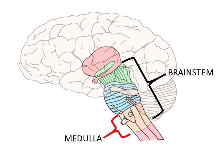
The Brain Stem
The posterior region on the top of the brain stem houses four small, rounded nuclei called the corpus quadrigemina. The upper two nuclei are the superior colliculi responsible for visual reflexes. Auditory reflexes produce the two lower nuclei called the inferior colliculi.

The reticular formation is a long, thin cluster of gray matter that spans most of the posterior brain stem. Consciousness consists of arousal and awareness. Arousal is the on/off switch of awareness. The reticular formation is essential for arousal, and its damage may lead to a coma.

The pons is in the middle of the brain stem on the anterior side. The pons acts as a bridge between the cerebellar hemispheres and the cerebellum and cerebrum. It also plays a role in respiration.
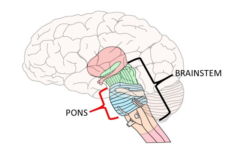
The medulla oblongata is at the base of the brain stem and connects to the spinal cord. The medulla oblongata is responsible for most of the body’s vital functions, such as heart rate, respiration, and blood pressure.