Muscle Tissue
Everything that moves in your body moves via one or more of the following processes: passive transport, active transport, cilia or flagella, gravity, or muscle contraction. Molecular motion occurs via active transport and passive transport. Body structures and large quantities of solutions move via muscle contraction and gravity.
Muscles allow us to run, play an instrument, and blink. They keep us alive by moving blood through our arteries and veins and moving air into and out of our lungs. Muscles move stuff.
Muscle tissue is vascular because it takes a lot of energy and nutrients to keep them moving. They move things via the contractile force of the proteins actin and myosin.
Skeletal Muscle
Skeletal muscle moves the skeleton, hence its name. It moves the eyeballs, diaphragm, and facial tissue, too. It contracts when swallowing and relaxes during defecation.
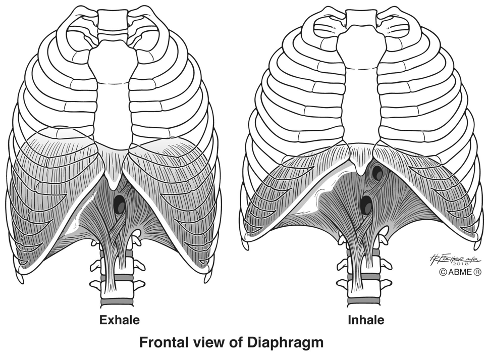
Skeletal muscle movements are both voluntary and involuntary. You can consciously control your diaphragm (it’s a skeletal muscle) when holding your breath. But breathing is an unconscious action as well.
A myocyte (muscle cell) or muscle fiber is a long cell with a fibrous appearance. Skeletal muscle fibers are multinucleated because they are the product of multiple cells fusing during prenatal development. Muscle fibers have striations or light and dark bands formed by the contractile proteins actin and myosin. During muscle contraction, myosin pulls on actin, which decreases the length of the cell. The shortening of the myocyte moves a bone.

Here is what skeletal muscle looks like under the microscope:


We are born with a fixed number of skeletal muscle fibers, which remain unchanged throughout postnatal growth. Our muscles increase in size during puberty, but the increase in size is because of an increase in actin and myosin within the muscle fibers, not more myocytes.
Skeletal muscle fibers are some of the longest-lived cells in the body, with an average 15-year lifespan. Muscle fiber replacement occurs when they die or are damaged beyond repair. Scattered throughout a muscle are satellite cells that repair or replace myocytes. But, if too many cells die at once or muscle tissue is severely injured, scar tissue replaces it. At 25, the rate of muscle fiber death exceeds regeneration leading to a decline in myocytes with age.
Cardiac Muscle
Cardiac muscle is in the walls surrounding the ventricles (heart chambers that pump blood) and, to a lesser extent, in the atria’ walls (heart chambers that receive blood from the veins). Cardiac muscle has striations, though they are fainter compared to skeletal fibers, and each cell has a single nucleus.
Here is what cardiac muscle looks like under the microscope:


Separating each cardiac fiber is an intercalated disk. The intercalated discs contain gap junctions, which results in a rapid connection between fibers. (A gap junction is a channel between two cells that speeds up communication.) The rapid spread of the electrical current produces a wavelike contraction along the myocardium so that the heart contracts as a unit.

Heart rate is under the control of the autonomic nervous system, so you have no conscious control over your heart rate. Two primary hormones raise or lower heart rate. Exposure to norepinephrine raises the heart rate, and exposure to acetylcholine lowers the heart rate.
Cardiac muscle fibers form during prenatal development. We are born with all the cardiac myocytes we will ever have because they do not regenerate. Therefore, when a cardiac muscle fiber dies, scar tissue replaces it, which weakens the heart.
Smooth Muscle
The autonomic nervous system controls smooth muscle contraction, so its contractions are involuntary. The parallel arrangement of actin and myosin in skeletal muscle and cardiac muscle causes the tissues to lengthen and shorten during muscle contraction and relaxation. However, smooth muscle does not have striations because the actin and myosin filaments are in a diagonal pattern, which gives it its smooth appearance. The oblique arrangement of actin and myosin pulls the smooth muscle fibers in multiple directions, causing them to ball up and squeeze hollow organs.

Here is what smooth muscle looks like under the microscope:


Smooth muscle is in the wall that surrounds most of the digestive tract. When it contracts, it compresses the GI tract by pushing food and water forward via strong wave-like contractions called peristalsis. And smooth muscle is in the walls surrounding the veins and arteries, which aids blood flow.

You probably woke up this morning thinking, “What does peristalsis look like in a living human being.” Well, here’s your answer:
Nervous Tissue
Nervous tissue forms the brain, spinal cord, and peripheral nerves (all the nerves outside the brain and spinal cord). Nervous tissue is vascular and comprises two major classes of cells: neurons and neuroglia.
Here is what nervous tissue looks like under the microscope:


Here are some fun facts about neurons and neuroglia:
- Neurons communicate with cells via electrical and chemical signals and receive sensory information from the external and internal environment.
- Neurons are the longest cell in the body, with some neurons being over two meters.
- Neurons develop in vitro and, to a lesser extent, during the first few months after birth. However, we make new olfactory neurons in the nasal cavity and neurons in the hippocampus (an area of the brain involved in learning and memory) daily.
- Neuroglia are the supporting cells of nervous tissue. Neuroglia help, repair, bathe, insulate, and protect neurons.

Key Concepts
- The primary function of muscle tissue is to move stuff.
- All muscle tissue types contain the contractile proteins actin and myosin.
- Skeletal muscle and cardiac muscle contain sarcomeres, the contractile units of a muscle fiber.
- The arrangement of actin and myosin in sarcomeres produces striations.
- Smooth muscle does not have striations due to the arrangement of actin and myosin.
- The brain, spinal cord, and peripheral nerves comprise nervous tissue.
- Neurons are the functional cell within nervous tissue.
- Neuroglia are the supporting cells within nervous tissue.