To understand how an organ works, you first need to know how the cells make it absorb, secrete, and excrete substances. The cell membrane controls molecule movement in and out of cells. The cell membrane is the gatekeeper to cells and the organs they form.
Water-Soluble and Lipid-Soluble Substances
Water-soluble and lipid-soluble are the two major substances that move in and out of cells. Ions, such as Na+, and polar molecules, such as glucose, are water-soluble. Water is a polar molecule with both a positive and negative changed pole. Water’s polarity allows it to form hydrogen bonds with ions, itself, and other polar molecules. For instance, the negative end of water forms a hydrogen bond with Na+, and the positive end forms a hydrogen bond with Cl–—i.e., how water separates NaCl into Na+ and Cl– ions. However, polar molecules and ions are not soluble in lipids like oil. Lipid-soluble, or nonpolar, molecules can dissolve in other nonpolar substances but not water. Nonpolar molecules do not have a charge, so water cannot form a hydrogen bond. The lack of bonding is why water (polar) and oil (nonpolar) do not make up a solution. A substance’s water or lipid solubility determines how it can or can’t enter or leave a cell.
| Ions | Polar Molecules | Nonpolar Molecules |
|---|---|---|
| Water-soluble | Water-soluble | Lipid-soluble |
| A positive OR negative charge | Positive end and negative end | No charge |
| Na+, Cl–, Ca2+, K+, HCO3– | 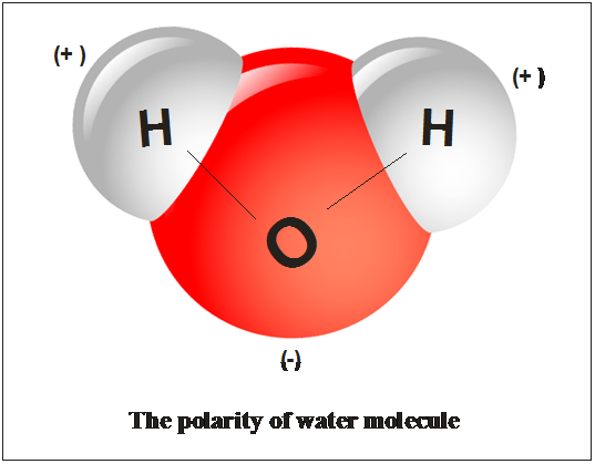 |  |
The Phospholipid Bilayer (aka Plasma Membrane)
Most of the cell membrane comprises phospholipids. A phospholipid is a lipid with a hydrophilic or water-liking end (polar head) and a hydrophobic or water-hating end (nonpolar tails). The intracellular and extracellular fluids are water-based solutions that attract the polar heads and repel the nonpolar tails. If cell membranes consisted of a single layer of phospholipids, the cell would form a micelle. A micelle is a ball of phospholipids where the polar heads face the extracellular fluid, and the nonpolar tails make up the ball’s internal portion. Since the nonpolar tails repel water, there would be no intracellular fluid. Therefore, the cell membrane forms a bilayer where the polar heads face the intracellular and extracellular fluids. The nonpolar tails comprise the middle of the membrane. The structure of the phospholipid bilayer regulates what can enter and leave a cell.


The phospholipid bilayer is a dynamic membrane because the phospholipids are always in flux. Nonpolar molecules can move across the plasma membrane via simple diffusion (See figure to the right) because they can squeeze through the polar heads and move within the nonpolar tails. Polar molecules cannot diffuse across the lipid bilayer because they cannot move through the layer of nonpolar tails. (No wall is impenetrable to something it tries to keep in and out, and the cell membrane is no exception. Water and specific ions can sneak across the phospholipid bilayer; however, only a fraction do this, and some cell membranes are more accessible than others.)

Proteins Floating in a Sea of Phospholipids
Protein doors are embedded in the cell membrane, allowing charged solutes to move in and out of cells. When charged solutes (except water) move passively through the protein’s door, it is facilitated diffusion. Aquaporins are protein doors that only allow water to move through the plasma membrane via osmosis.
A wide range of protein types floats around in the plasma membrane. Each type of protein has a specific function. Without plasma proteins, the cell’s physiology would differ from its current form.

Receptor proteins have a groove where ligands (hormones and neurotransmitters) with complementary shapes can attach. A ligand binds to a receptor protein and starts a cellular response. For example, insulin receptors are abundant in skeletal muscle fibers’ membranes since muscle requires a lot of glucose to function. Since glucose is polar and cannot diffuse through the phospholipid bilayer, it must move in via a door (channel protein). The problem is a locked door that requires a hormone to unlock it. When the hormone insulin binds to its receptor protein, it starts a cellular response, ending with a channel protein (door) opening and glucose entering the cell.
Transport proteins use active transport (cell uses energy) or passive transport (cell does not use energy) to move ions and polar molecules through the cell membrane. There are two types: (1) carrier proteins move more substantial quantities of solutes across the membrane via passive or active transport, and (2) channel proteins use passive transport to move smaller ions and polar molecules through the membrane.
The primary carrier proteins we will focus on this year are protein pumps. For example, the sodium-potassium pump uses active transport to pump Na+ and K+ across the plasma membrane. Pumping Na+ and K+ maintains the proper ratio of ions inside and outside a neuron.
Channel proteins are a diverse group that requires different stimuli to open and close.
Voltage-gated channel proteins open and close because of a change in charge along the cell membrane. Neurons have Na+ and K+ voltage-gated channel proteins within their plasma membrane that opens in response to an electrical current. The plasma membrane of a neuron has K+ channel proteins that are always open where K+ can diffuse back and forth along the membrane.
Mechanical-gated channel proteins open in response to pressure, such as touch or vibrations.
Ligand-gated channel protein is what would be the result if a receptor protein and a channel protein had a baby. A ligand attaches to the protein’s receptor site, which causes the channel to open and ions or polar molecules to move in or out of the cell.
Cell adhesion proteins in the phospholipid bilayer bind cells together via anchorage dependence. Different cell adhesion proteins exist, but we will focus on gap junctions only this school year.
Gap junctions form when a channel protein on one cell forms a bond with a channel protein on another, connecting the two cells’ cytoplasm. The connection allows for the rapid exchange of ions, molecules, and electrical impulses between the two cells. For example, the gap junctions between cardiac muscle fibers send electrical impulses from cell to cell, so the heart contracts as a unit.
Cells can distinguish your body’s cells from foreign cells via membrane proteins called self-antigens. Self-antigens are present on all your nucleated cells on a plasma protein called the major histocompatibility complex (MHC), which we will discuss in the Immunity Unit. Each person has a unique self-antigen, and your immune system will attack and destroy any cell with a different antigen. Genetics determines the shape of an individual’s self-antigen, so if you have an identical twin, you share the same self-antigen.

Passive Transport
Passive transport is the process in which a substance moves down its concentration gradient. Ions and compounds want to move from high to low concentrations until they reach equilibrium. A cell uses passive transport to gain nutrients and water and eliminate waste or an overabundance of nutrients. The data table below defines and gives examples of different passive transports.

Active Transport
Sometimes a cell needs more of something that moves against the substances’ concentration gradient, or the cell needs to get rid of something too large to fit through a carrier protein. Gaining substances against their concentration gradient or getting rid of big stuff requires a cell to use energy via active transport.
