What is a Neuron?
The principal cell of the nervous system is the neuron. Or what I like to call it, a nerdon.
Stop. Just stop.
There are about 86 billion neurons in your brain, which form over 100 trillion connections. There are also neurons in your spinal cord and nerves throughout the body, though they are fewer in number. Neurons receive and transmit electric impulses called action potentials and graded potentials. There is a gap between most neurons and the cell they communicate with called the synaptic cleft. The electrical impulses cannot jump the synaptic cleft, so neurons release ligands called neurotransmitters. The neurotransmitters diffuse across the synaptic cleft and attach to receptor proteins on the postsynaptic cell. The message from the neurotransmitter will either excite or inhibit the postsynaptic cell.
Neuron Anatomy
Most neurons have the following structures:
- Dendrites
- Short branches of a neuron that carry impulses TOWARDS the cell body (soma)
- Receive stimuli from another cell or a chemical, light, or sound
- Neurons may have one or more dendrites
- Usually have many divisions called dendritic branches
- Soma (Cell Body)
- Processes information collected from the dendrites
- Site of neurotransmitter synthesis
- Axon Hillock (“The Trigger Zone”)
- A swelling of the soma where the axon begins
- The “trigger” zone for most nerve impulses (known as action potentials)
- If neurons were Nerf guns, this would be a trigger
- Axon
- The long extension of a neuron that carries the impulse (action potential) AWAY fromthe cell body (soma)
- All neurons have only ONE axon
- If neurons were Nerf guns, this would be a barrel
- Myelin sheath
- A fatty, segmented covering wrapped around axons, which increases the speed of nerve impulses
- The more myelin surrounding an axon, the faster the nerve impulse
- Myelin surrounds most axons (we are born with very few myelinated axons
- Myelin only surrounds axons; there is no myelin on dendrites
- Nodes of Ranvier
- Small gaps in the myelin sheath along the axon
- These gaps help move the nerve impulse down an axon
- Terminal Branches
- The branching segments that start at the distal end of an axon and stop at the terminal endings
- Terminal Endings
- Expanded end of the terminal branches that contain synaptic vesicles filled with neurotransmitters
- The site where the electrical nerve impulse (action potential) becomes a chemical impulse
- If a neuron were a Nerf gun, this would be where the Nerf dart (neurotransmitters)leaves the Nerf gun.

CNS and PNS
The central nervous system (CNS) comprises the brain and spinal cord. The peripheral nervous system (PNS) includes all the nervous tissue outside the brain and spinal cord – i.e., spinal nerves and cranial nerves.
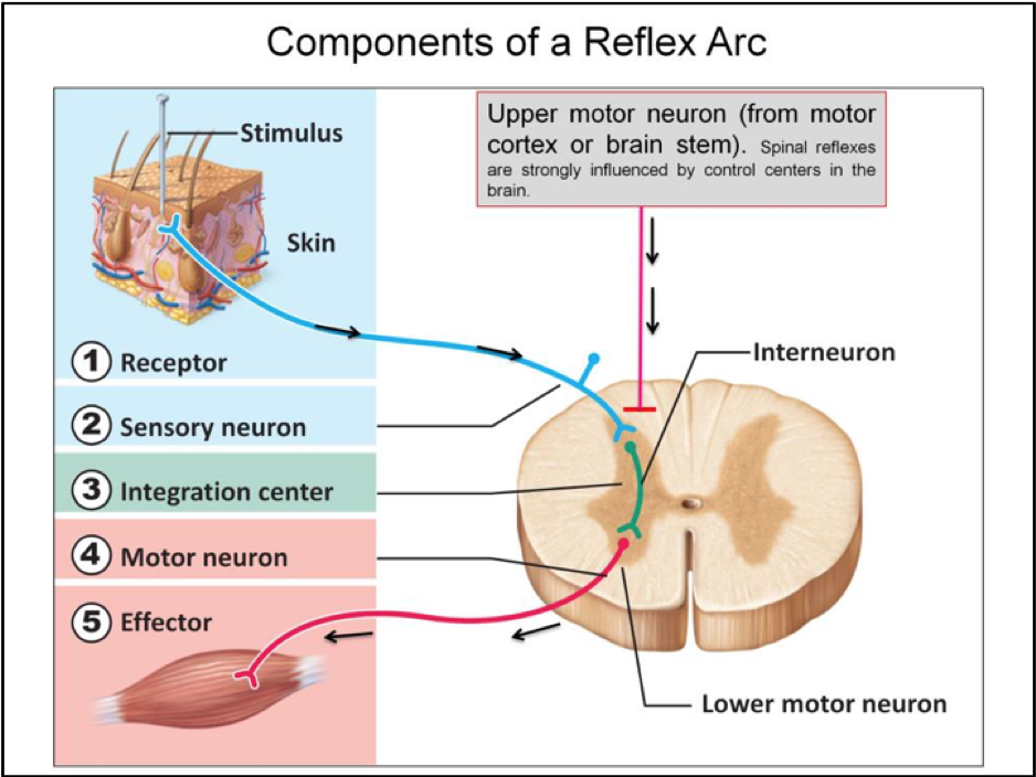
Sensory Neurons, Motor Neurons, and Association Neurons
Reading the words on this page requires sensory neurons, association neurons(interneurons), and motor neurons. Sensory neurons are afferent because they send sensory information to the central nervous system. As you look at the words on this page, photons in light stimulate the rods and cones (sensory neurons) in your retina. The rods and cones stimulate the sensory neurons of the optic nerve. Once the sensory information reaches your brain, association neurons analyze the data.
Association neurons (interneurons) are only in the central nervous system and connect sensory neurons to motor neurons. Data processing, analysis, and storage are the primary functions of association neurons. For example, your interneurons receive the color, depth, shape, time, and all the other components of sight from sensory neurons. Association neurons connect the pieces, which form letters. Letters are combined to form words, then grammar and syntax, followed by sentences. After analysis, association neurons form thoughts and memories that may translate into motor movement.
Motor neurons are efferent neurons because they send information away from the brain and spinal cord. The data traveling along motor neurons will tell the ocular muscles how to move when reading this chapter. Motor neurons send messages to muscles, glands, and organs.
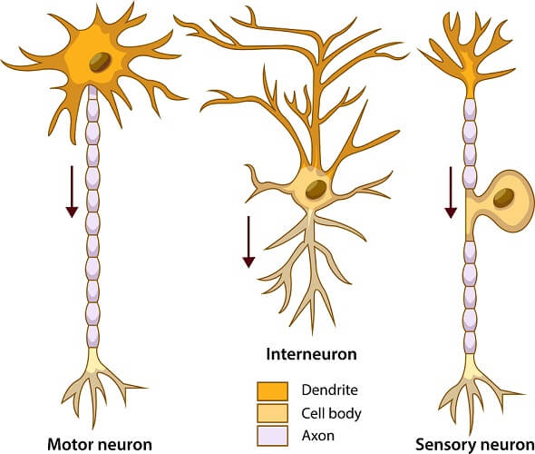
Unipolar, Bipolar, and Multipolar Neurons
The neuron type depends on how many dendrites it has. Most sensory neurons are unipolar, meaning they have only one long axon with no dendrites attached to the soma (or they may have no dendrites). Bipolar neurons are the rarest type of sensory neurons. The retina, olfactory neurons in the nasal cavity, and the auditory nerve consist of bipolar neurons.
All motor neurons and association neurons are multipolar neurons. Multipolar neurons have one axon and many dendrites. Dendrites attached to the cell body tend to be branching, like trees. One large dendrite can give rise to many smaller dendritic branches. For example, the Purkinje cells in the cerebral cortex can have more than 100,000 dendritic branches, and each division can form multiple connections with other neurons. The brain’s neural network is so vast that it has at least 100 trillion connections.
Gray Matter and White Matter
Gray matter refers to a gathering of neuron cell bodies. However, not all gray matter is gray. The red nucleus is a cluster of cell bodies (gray matter) that have a reddish color. White matter consists of clumps of myelinated axons. The two cerebral hemispheres in your brain connect via a large bunch of white matter. Without this connection, the two sides of the cerebrum can not communicate, resulting in cognitive impairments.

What are Neuroglia?
Neuroglia are cells in the nervous system that protect neurons, help with neural communication, and regulate blood flow to the brain. The number of neuroglia in the brain is under debate. Most textbooks state there are ten times more neuroglia than neurons, while recent studies have reduced the ratio to 3:1 and even 1:1. So, there are somewhere between 86 billion and 860 billion neuroglia in your brain.
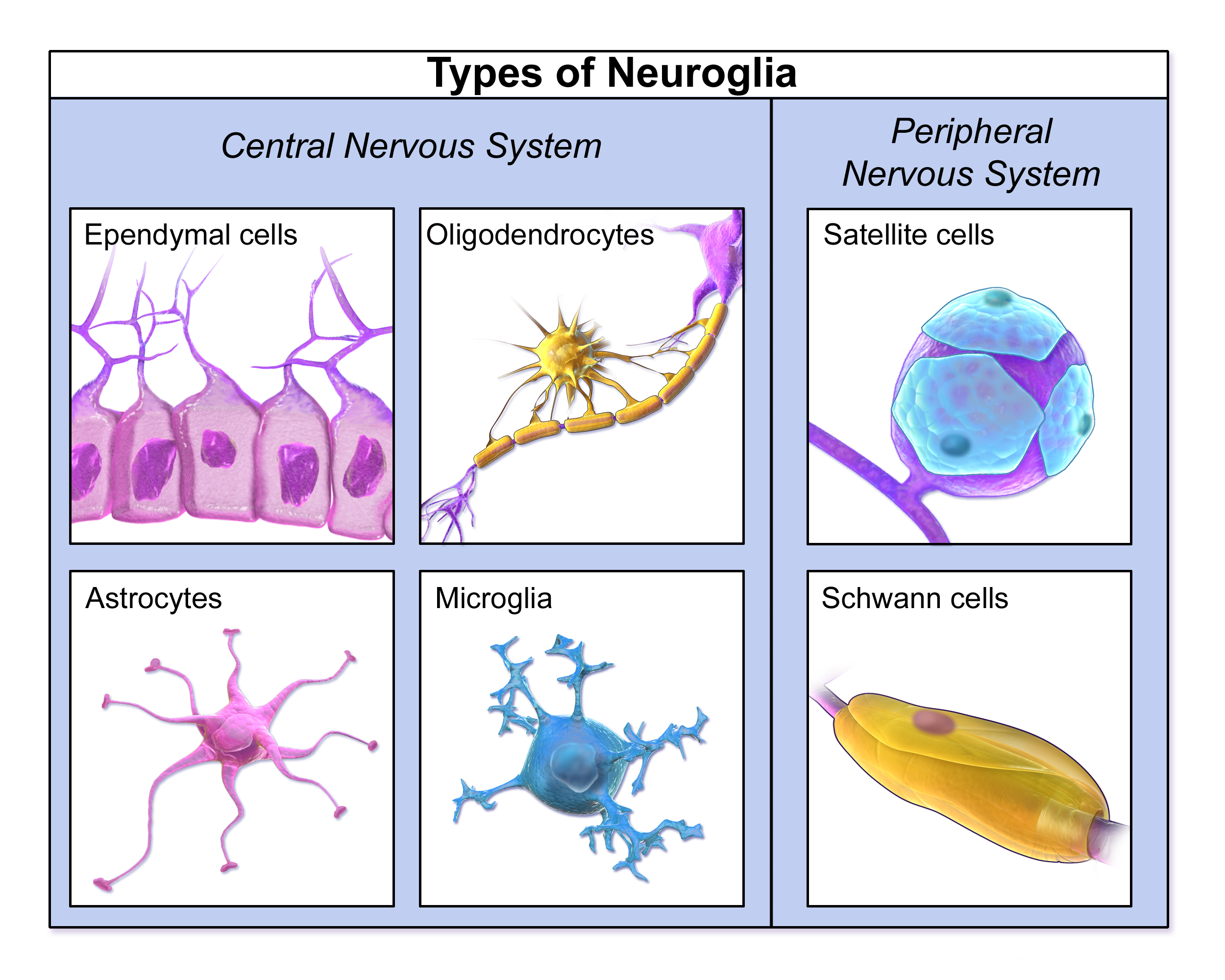
Postnatal neuroglia can undergo cell division, something almost all neurons cannot do. (A few neurons in the nasal cavity divide, and new neurons in the hippocampus form from stem cells through life.) Since neurons rarely divide, neuron cancers are rare, with most happening in utero. Most brain cancers originate from neuroglia (gliomas) or meningeal cells (cells of the membranes that surround the brain).
Glioblastomas are the most destructive and common type of brain cancer. After diagnosis, the median survival rate for glioblastoma is a year.
Brief Chapter Summary
- Neurons are the functional cells of the nervous system
- Dendrites receive information from other neurons, cells, or the environment
- A nerve impulse (action potential) travels down an axon
- A neuron can have many dendrites but has only one axon
- The myelin sheath insulates axons, speeding up nerve impulses
- Sensory neurons are afferent because they send information to the brain
- Motor neurons are efferent because they send information away from the brain